


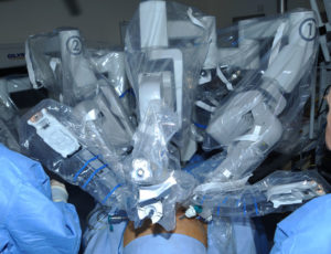


MRI-US Fusion Biopsy: Advancing Prostate Cancer Diagnosis
Prostate cancer remains one of the most common cancers in men, making accurate and early diagnosis a cornerstone of effective treatment. In recent years, MRI-US fusion biopsy has emerged as a transformative tool in the detection and management of prostate cancer. Combining the anatomical detail of magnetic resonance imaging (MRI) with the real-time functionality of ultrasound (US), this approach enhances diagnostic precision while minimising unnecessary biopsies.
What Is an MRI-US Fusion Biopsy?
MRI-US fusion biopsy is a targeted biopsy technique that leverages the strengths of two imaging modalities:
- Multiparametric MRI (mpMRI): This imaging technique captures detailed images of the prostate, highlighting areas suspicious for cancer using advanced sequences such as diffusion-weighted imaging (DWI), dynamic contrast-enhanced (DCE) imaging, and T2-weighted imaging. Lesions are scored on the PI-RADS (Prostate Imaging Reporting and Data System) scale to indicate their likelihood of malignancy.
- Ultrasound (US): While traditional transrectal ultrasound (TRUS) is used for random sampling of the prostate, its ability to delineate specific lesions is limited. Fusion biopsy technology integrates MRI-identified targets with real-time ultrasound, guiding precise sampling of suspicious areas.
How Does MRI-US Fusion Biopsy Work?
The process typically involves the following steps:
- Pre-Biopsy MRI: The patient undergoes a multiparametric MRI, which identifies and localises suspicious lesions within the prostate.
- Fusion Technology: Using specialised software, the MRI images are fused with real-time ultrasound during the biopsy procedure. This ensures the MRI-identified lesions are accurately targeted.
- Biopsy Procedure:
- Performed transrectally or transperineally, depending on patient and physician preference.
- Real-time ultrasound guides the biopsy needle, while the fusion system ensures precise sampling of the MRI-detected lesions.
- Systematic sampling may also be performed to assess the prostate comprehensively.
Benefits of MRI-US Fusion Biopsy
MRI-US fusion biopsy offers several advantages over traditional methods:
- Improved Cancer Detection:
- Targets suspicious areas with high precision, improving the detection of clinically significant cancers.
- Reduces the likelihood of missing aggressive cancers that may not be sampled during random biopsies.
- Fewer Unnecessary Biopsies:
- Helps avoid sampling benign areas unnecessarily, reducing the risks and discomfort associated with over-biopsy.
- Better Risk Stratification:
- Provides more accurate information about tumour location, size, and grade, enabling tailored treatment planning.
- Minimised Side Effects:
- Lower risk of complications such as bleeding, infection, and urinary retention due to reduced sampling.
Who Should Consider an MRI-US Fusion Biopsy?
This technique is particularly beneficial for:
- Men with a raised PSA level and a prior negative biopsy, but persistent suspicion of cancer.
- Patients with a PI-RADS 3, 4, or 5 lesion on MRI, indicative of moderate to high suspicion of cancer.
- Those requiring active surveillance to monitor low-risk prostate cancer, where precise sampling is essential for tracking disease progression.
Limitations of MRI-US Fusion Biopsy
While MRI-US fusion biopsy represents a significant advancement, it is not without limitations:
- Access and Cost: The need for advanced imaging and software may limit availability in some settings.
- Technical Expertise: The procedure requires specialised training for radiologists and urologists to interpret MRI findings and perform the biopsy effectively.
- False Negatives: Although rare, some cancers may remain undetected, particularly in areas of the prostate that are challenging to visualise with MRI.
The Future of Prostate Biopsy
As imaging technologies continue to evolve, the role of MRI-US fusion biopsy in prostate cancer diagnosis is expected to expand. Ongoing developments in artificial intelligence (AI) and machine learning are enhancing lesion detection and PI-RADS scoring, making the fusion process even more precise. Additionally, integrating genomic testing with biopsy results offers the potential for personalised, risk-adapted treatment strategies.
Conclusion
MRI-US fusion biopsy has revolutionised the way prostate cancer is diagnosed, offering unparalleled accuracy and reducing the burden of unnecessary procedures. For men with suspected or known prostate cancer, this innovative approach represents a step forward in achieving better outcomes and improved quality of life.
If you have concerns about prostate health or are considering a biopsy, consult with your urologist to determine if MRI-US fusion biopsy is the right choice for you.